The nose, occupying a prominent position on the face, comprises several distinct subunits, with the nasal alar being particularly delicate and distinctive. Together with other subunits, the nasal alar contributes to the overall contours of the nose, encompassing both convex and concave shapes.
Alar defects can arise from a variety of factors, including trauma, scars, tumor removal, congenital deformities, and, more recently, complications associated with filler injections in the nasolabial fold, which can lead to nasal alar necrosis and subsequent defects.
In countries like China, nasal alar defects are predominantly caused by industrial trauma rather than surgical procedures like Mohs micrographic surgery. These traumatic defects often involve the alar cartilages, crucial structures that provide support to the nose.
Unlike Caucasians, Asians typically have weaker alar cartilages, which necessitates careful evaluation of their involvement in assessing the depth of the defect. Furthermore, Asian patients are prone to hypertrophic scar formation following trauma or surgery, requiring careful consideration of scar formation as a potential postoperative complication.
In a recent study published in the Chinese Journal of Plastic and Reconstructive Surgery, a team of researchers from China introduced an accurate classification system and systematic treatment algorithm for nasal alar defects in Asian populations.
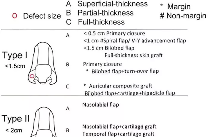
Corresponding author of the study, Professor Danru Wang, a specialist in plastic and reconstructive surgery at the Shanghai Ninth Hospital, elaborates on the significance of their work. “Based on the defect classification system and clinical expertise, we have developed a treatment algorithm that offers tailored approaches for different types of alar defects.”
The classification system divides alar defects into three main types: Type I for small defects (diameter < 1.5 cm), Type II for medium-size defects (diameter between 1.5 and 2 cm), and Type III for large defects (diameter > 2 cm). Each type is further subdivided based on the thickness of the defect: superficial thickness (Type A), partial thickness (Type B), and full thickness (Type C). Additionally, Type IV defects indicate combined defects involving other subunits such as the cheek or maxilla.
This algorithm provides a valuable framework for assessing the severity of the deformity, while the accompanying reconstructive algorithm assists in selecting the most suitable surgical approach for each type of defect.