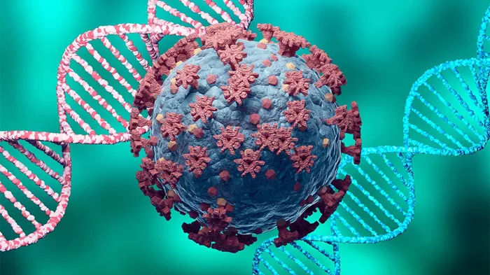
Recent research has provided critical insights into the unique ways in which SARS-CoV-2 evolves within the central nervous system (CNS), revealing significant implications for understanding the virus’s behavior and potential neurological complications. The study, published in Nature Microbiology, compares the evolutionary patterns of SARS-CoV-2 in the lungs and the CNS, highlighting differences in viral diversification that could explain the severe neurological symptoms associated with COVID-19.
COVID-19 Pathology: Beyond the Lungs
SARS-CoV-2, the virus responsible for the COVID-19 pandemic, primarily targets lung epithelial cells. However, it can also infiltrate the CNS, leading to acute kidney injury, myocarditis, thromboembolism, and potentially severe neurological complications. The precise mechanisms through which SARS-CoV-2 causes these diverse pathologies remain incompletely understood.
The viral spike glycoprotein (S), which facilitates the entry of SARS-CoV-2 into host cells, plays a critical role in this process. The S protein comprises two subunits, S1 and S2, at the furin cleavage site (FCS). Over time, SARS-CoV-2 has evolved to produce more infectious variants, with mutations in the FCS influencing how the virus interacts with host cells.
While many studies have focused on the viral load and mutations within the lungs, the dynamics of these mutations in other tissues, such as the brain, remain unclear. This study seeks to bridge that gap by exploring how the virus evolves differently in various host tissues.
Study Overview
Researchers employed two distinct mouse models to investigate how SARS-CoV-2 evolves in different tissues and whether pre-existing immunity affects this evolution. The mice were divided into experimental groups and vaccinated either intranasally or intracranially with adenovirus vector vaccines encoding either the SARS-CoV-2 S protein (Ad5-S) or the nucleocapsid protein (Ad5-N). A phosphate-buffered saline (PBS) solution was used as a control. After three weeks, the mice were exposed to a high frequency of mutations in the spike FCS.
Focus-forming assays, real-time quantitative polymerase chain reaction (RT-qPCR), and whole-genome sequencing were employed to analyze viral evolution. Researchers also calculated Shannon entropy to assess intra-host viral diversification across different tissues and animals.
Key Findings
The study revealed that SARS-CoV-2 strains lacking the FCS exhibited reduced virulence in the lungs, likely due to an increased reliance on the TMPRSS2-independent endosomal entry pathway. Interestingly, these FCS-deficient variants showed a greater propensity to infect CNS tissues, mirroring findings from previous research on other coronaviruses.
Notably, the FCS-deficient pseudovirus demonstrated a diminished ability to enter lung cells compared to visceral adipose tissue (VAT) cells. This reduced efficiency was linked to lower viral titers and decreased pathology in the lungs following intranasal inoculation.
Vaccination status and formulation did not significantly impact viral divergence in the lungs. However, viral diversity in brain isolates varied significantly, regardless of the type of vaccination. In some vaccinated mice, viral diversity was higher in the lungs, while in others, the brain exhibited greater diversity.
Among the vaccinated groups, mice that received the Ad5-S vaccine showed reduced viral diversity in the lungs but maintained higher diversity in the brain. This finding suggests that while the Ad5-S vaccine may limit viral evolution in the lungs, the virus continues to diversify within the CNS.
A notable enrichment in S protein diversity, particularly around the FCS, was observed. This finding aligns with previous studies indicating that neuroinvasion triggers selective pressure for mutations or deletions in the FCS, regardless of prior immunity status.
Implications and Future Research
The study’s findings underscore the complex dynamics of SARS-CoV-2 evolution within different tissues and the role of selective pressure in shaping viral behavior. The observed rapid proliferation of the FCS mutant strain in neuronal cells suggests that the CNS environment may exert unique selective pressures on the virus, potentially driving the emergence of new variants.
These insights are crucial for understanding the neurological complications associated with both acute COVID-19 and long COVID. The study highlights the need for further research to explore the role of compartmentalization in the evolution of SARS-CoV-2 and the potential for direct viral infection of the CNS to contribute to neurological symptoms.
As the world continues to grapple with the impacts of COVID-19, this research provides a critical foundation for developing more targeted therapies and vaccines that address the diverse ways in which SARS-CoV-2 can affect the human body.
[inline_related_posts title=”You Might Be Interested In” title_align=”left” style=”list” number=”6″ align=”none” ids=”3341,773,3343″ by=”categories” orderby=”rand” order=”DESC” hide_thumb=”no” thumb_right=”no” views=”no” date=”yes” grid_columns=”2″ post_type=”” tax=””]


