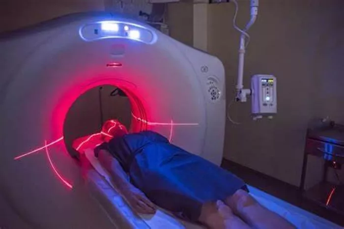
Researchers at St. Jude Children’s Research Hospital have made a significant breakthrough in the diagnosis of neurological diseases, enhancing the capabilities of positron emission tomography (PET) imaging. The team repurposed edaravone, an antioxidant used in the treatment of amyotrophic lateral sclerosis (ALS), as a probe to detect oxidative stress in the brain. This advancement, detailed in a study published in Nature Biomedical Engineering, holds promise for earlier diagnosis of conditions such as ALS and Alzheimer’s disease.
Oxidative stress, caused by an accumulation of reactive oxygen and nitrogen species (RONS), is a key factor in the progression of various neurodegenerative diseases. These reactive molecules, while essential for normal cellular functions, can lead to cell damage when they accumulate in excess. The ability to detect oxidative stress through noninvasive imaging techniques could allow for the earlier detection of diseases like ALS and Alzheimer’s, improving the chances for effective treatment before irreversible damage occurs.
According to the research team, oxidative stress is not only a hallmark of neurodegenerative diseases but also plays a role in other neurological conditions such as stroke. In stroke, the primary injury is compounded by a secondary immune response, which includes a burst of reactive oxygen and nitrogen species. This “oxidative burst” can cause extensive damage to brain tissue, making it a critical target for early diagnosis and intervention.
Kiel Neumann, PhD, the study’s corresponding author and a member of the St. Jude Department of Radiology, explained the importance of understanding oxidative stress in neurological diseases: “The harm in these conditions often comes from the immune response, which releases a cascade of oxidative chemicals. Detecting this burst of oxidative stress is key to understanding the damage that is being done.”
In an innovative approach, Neumann and his team radiolabeled edaravone, replacing atoms in the molecule with radioactive isotopes to create a new imaging tool. The radiolabeled edaravone, when administered to patients, emits positrons that can be detected by PET scans, highlighting areas of the brain where oxidative stress has accumulated. This process allows for a detailed view of the brain’s response to oxidative damage, enabling earlier detection of diseases that were traditionally diagnosed only after significant physical symptoms appeared.
“The goal in imaging is to create contrast, so you want a substance that interacts rapidly with its target and clears quickly, allowing for immediate visualization,” Neumann said. “What makes edaravone unique is that it undergoes a structural and polarity change when it reacts with oxidative stress, allowing it to remain in the affected cells and provide clearer imaging contrast.”
This new technique not only enhances diagnostic capabilities but also allows edaravone to retain its role as an antioxidant treatment, making it a potential dual-purpose tool for both diagnosis and therapy. The researchers have found that only nanogram to microgram amounts of the drug are needed for imaging, which are small enough that the body does not detect its presence, minimizing side effects.
Neumann emphasized the broader impact of this research: “Ultimately, our goal is to integrate this technology into clinical care to aid in the management of neurological diseases. Early detection and therapeutic intervention are the future of disease management.”
This groundbreaking advancement marks a critical step toward improving the diagnosis and treatment of brain diseases, offering new hope for patients and paving the way for further innovations in medical imaging and treatment.
Related Topics